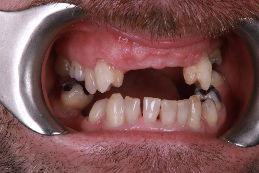
Anthony presented with a partially edentulous maxilla and a failing lower right posterior segment. The case was selected and presented as a live surgery demonstration in front of over 1,000 clinicians at the European Symposium for Osteem, showcasing immediate digital workflows, FP1 planning, and soft tissue management in real time.
The upper arch presented with a mix of stable and failing teeth.
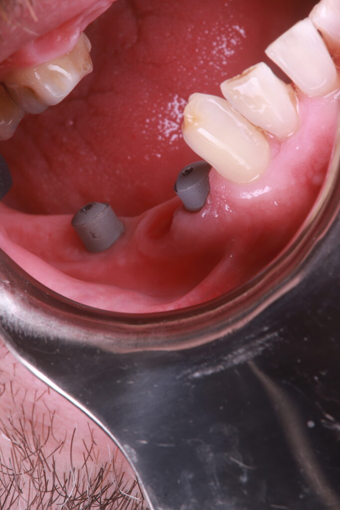
Only UR4, UR5, and UL4 were extracted due to mobility and poor prognosis. In the mandible, LR6 was removed and replaced with a short-span implant bridge.
This case forms part of an advanced training portfolio in FP1 prosthetics, and will be used for ongoing collaboration in digital and restorative education.
Diagnosis and Treatment Plan
Maxillary Arch
Diagnosis: Partially edentulous upper arch with anterior tooth loss and posterior mobility
Treatment:
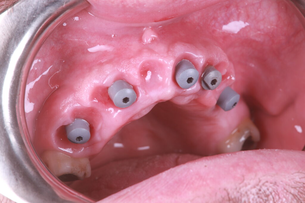
Extraction of UR4, UR5, UL4
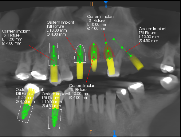
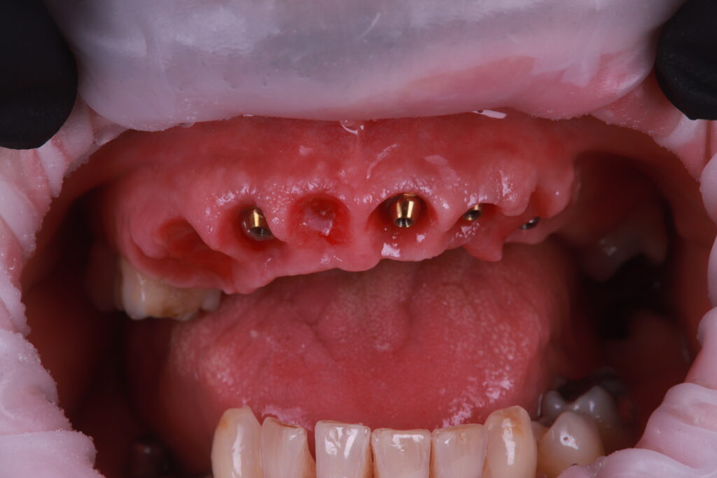
Placement of five implants in the anterior and premolar regions
Delivery of immediate 3D-printed provisional prosthesis
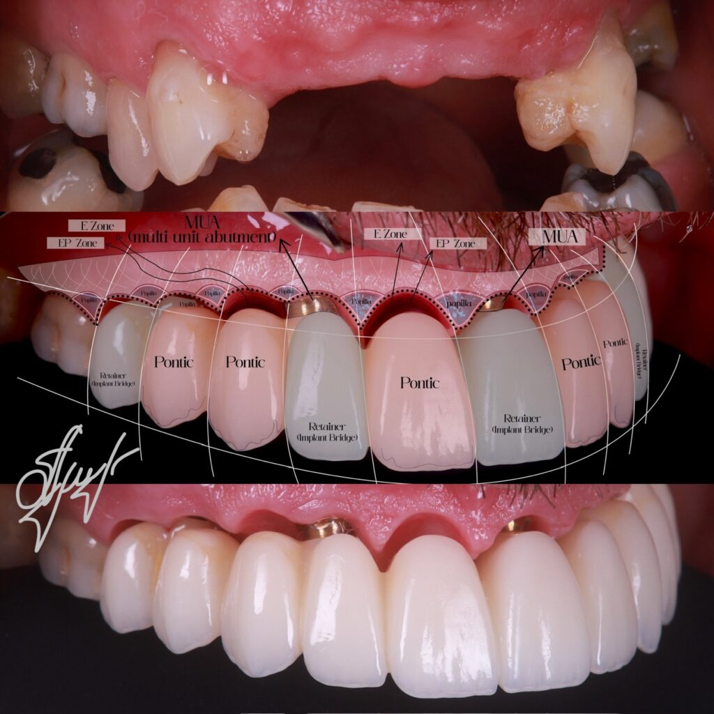
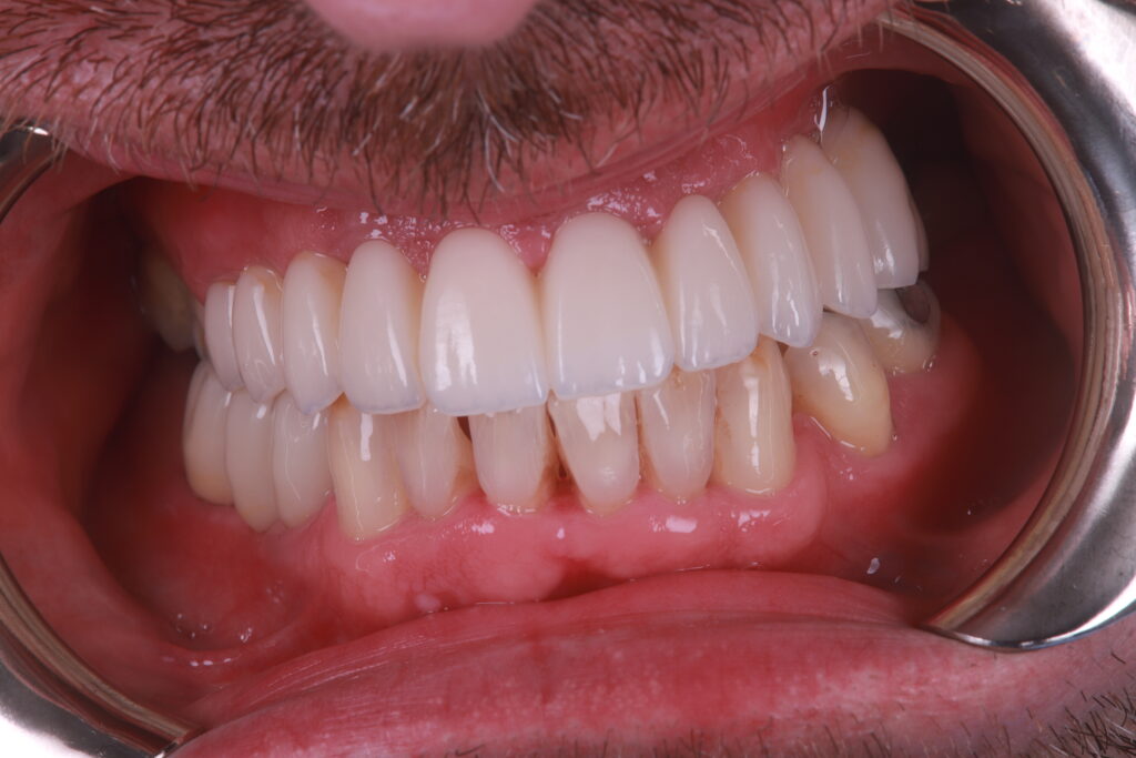
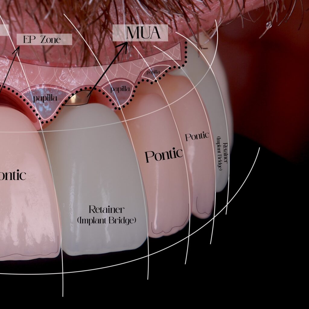
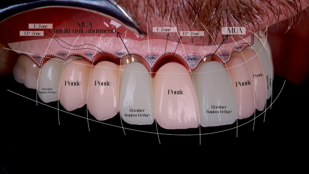
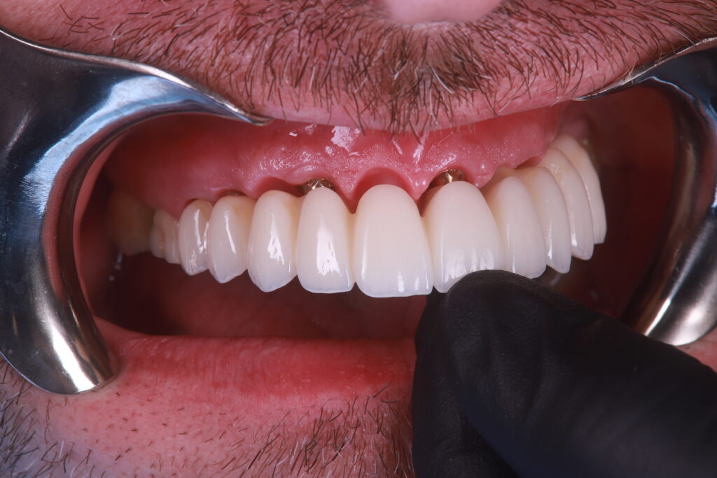
Final restoration with FP1 monolithic zirconia prosthesis, supported by existing and new implants
Maintenance of posterior teeth to preserve bone and function
Mandibular Arch
Diagnosis: Failing LR6 with posterior occlusal deficiency
Treatment:
Extraction of LR6
Placement of two implants and restoration with a 3-unit posterior bridge
Materials and Techniques Used
Implants: Placed using guided surgery with intraoperative imaging
Digital Workflow:
iTero for intraoral scanning and soft tissue capture
Photogrammetry 2.3D for precise implant position registration
Same-day 3D printing of provisional prosthesis
Restorations:
FP1 zirconia prosthesis (upper)
Same-day 3D printing of provisional prosthesis
intraoral scanning and soft tissue capture
Full soft tissue and implant position capture
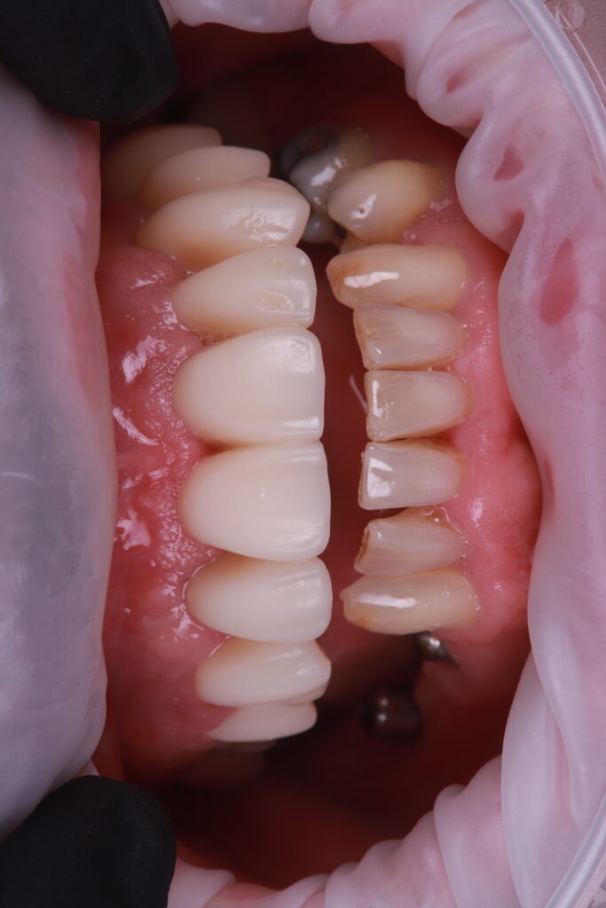
Final Provisional
2 weeks post-op
Outcome
This case illustrates how immediate digital integration, soft tissue control, and implant-guided prosthetic planning can be used to deliver an FP1 zirconia prosthesis with long-term aesthetic and functional stability.
The live procedure included:
Same-day guided surgery
3D-printed provisional prosthesis delivered within hours
Full soft tissue and implant position capture using iTero scanning and photogrammetry
Over a 6-month provisional phase, soft tissue architecture was carefully matured to support the final FP1 prosthesis. The patient achieved an excellent aesthetic result, with healthy, maintainable soft tissue contours and a balanced occlusion.